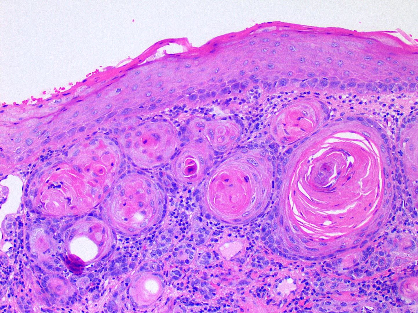
Gambaran khas SCC pada frozen section dan histopatologi
- > "Typical SCC has nests of squamous epithelial cells arising from the epidermis and extending into the dermis . The malignant cells are often large with abundant eosinophilic cytoplasm and a large, often vesicular, nucleus. Variable keratinisation (keratin pearls etc) is present ."
secara umum SCC bersarang pada sel epitel dari epidermis yang meluas ke dermis. Malignan sel sering muncul secara berlimpah dengan sitoplasma yg eosinofilik yang besar, sering vesikular dan nukleas. Keratin pearls dapat muncul. Namun SCC memiliki banyak varian yang memunculkan banyak jenis penampakan histopatologis antara lain: spindle cell SCC, clear cell SCC,desmoplastic SCC,dsb
"histopathologically, single-cell SCC is composed almost entirely of singular atypical cells, which exist either individually or loosely arranged as nests in the dermis. There is an overall lack of cohesiveness amongst atypical cells, and usually no connection to the overlying epidermis or adnexal structures."
Secara histopatologi , Single cell SCC terdiridari sel2 singular yang atipikal yang menyebar ke dermis dan memiliki karaktersitik kekurangan kohesiveness (menyebar/tidak mengumpul) dan biasanya tidak terhubung dengan epidermis diatasnya.
Sumber:
- Valerie R. Yanofsky, Stephen E. Mercer, Robert G. Phelps, "Histopathological Variants of Cutaneous Squamous Cell Carcinoma: A Review", Journal of Skin Cancer, vol. 2011, Article ID 210813, 13 pages, 2011. https://doi.org/10.1155/2011/210813
- Dermznet.com
- Weedon D. Skin pathology, Third Edition. Elseiver Ltd. 2010; Squamous cell carcinoma p.691–700